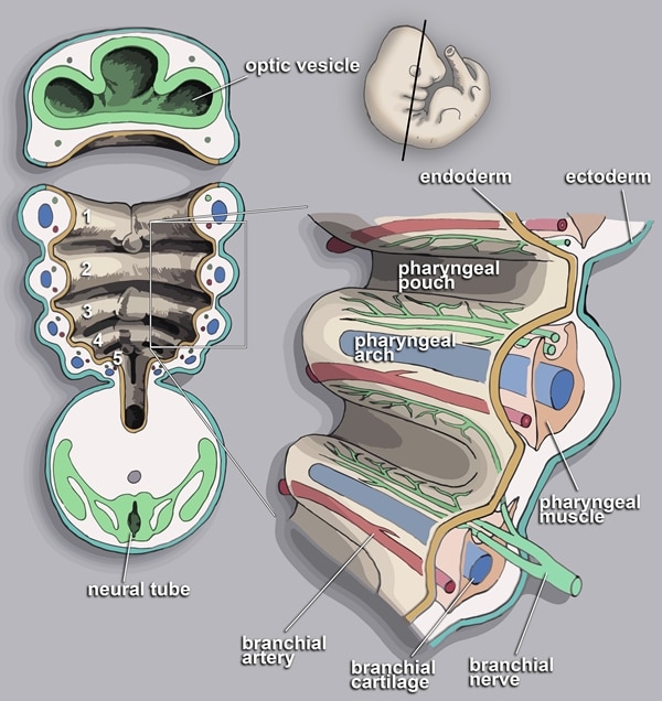
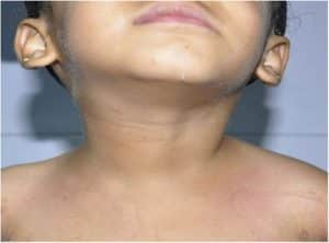
An abranchial cleft cyst is a cyst as a swelling in the lateral part of the neck near the sternocleidomastoid muscle. Sometimes, the branchial cleft cyst can occur with an opening known as the fistula. The cause is usually due to a birth defect arising from the failure of fusion of the second and third branchial arches. The branchial arches are related to gill slits. In fish and amphibians, these structures are responsible for the development of the gills, hence the name branchial (branchia is Greek for gills).
Although the second branchial cleft is the most common area, the cysts can also less commonly develop from the first, third, or fourth clefts.
Treatment options include conservative (no treatment) and branchial cleft cyst surgery.

The pharyngeal arches as seen during embryonic development
More: History of Surgery
Symptoms of Branchial Cleft Cyst
Most branchial cleft cysts present in late childhood or early adulthood as a solitary, painless mass, which went previously unnoticed, that has now become infected (typically after an upper respiratory tract infection). Fistulas, if present, are asymptomatic until infection arises.
Diagnosis of Branchial Cleft Cyst
The branchial cleft cyst can be diagnosed through several medical examinations. More often, a physical examination is sufficient to diagnose a branchial cleft cyst but a tissue biopsy or imaging may be required especially for masses presenting in adulthood which should be considered cancerous until proven otherwise.
Some forms of cancer such as carcinomas of the tonsil, tongue base and thyroid may all present as cystic masses of the neck. It is therefore pertinent to conduct confirmatory medical tests to ascertain the nature of the cyst.

Treatment of Branchial Cleft Cyst
There are two options for the treatment of branchial cleft cyst namely; conservative (no treatment) and surgical excision. Usually, treatment of branchial cleft cyst is not necessary, unless they become painful or restrict movement. They are usually removed for cosmetic reasons if they grow very large or for histopathology to verify that they are not a more dangerous type of tumor such as a liposarcoma.
Branchial cleft cyst surgery is a surgical operation that is carried out to remove the cyst. Surgical excision is the definitive treatment for branchial cleft cysts. A series of horizontal incisions, known as a stairstep or stepladder incision, is made to fully dissect out the occasionally tortuous path of the branchial cleft cysts.
Branchial cleft cyst surgery is best delayed until the patient is at least age 3 months. Definitive branchial cleft cyst surgery should not be attempted during an episode of acute infection or if an abscess is present. Surgical incision and drainage of abscesses are indicated if present, usually along with concurrent antimicrobial therapy.
The traditional surgical approach has the main downfall of relatively significant scarring. Alternatives to the open surgical method have been proposed, including a retro auricular approach, a facelift approach, and endoscopic-assisted removal. All of the newer surgical methods may be limited in the full visualization of the lesion. A recent case-controlled study suggested that an endoscopic retroauricular approach may provide good surgical clearing with minimal scarring for second branchial cleft cysts.
Following branchial cleft cyst surgery, recurrence is common, usually due to incomplete excision. Often, the tracts of the cyst will pass near important structures, such as the internal jugular vein, carotid artery, or facial nerve, making complete excision impractical due to the high risk of complications.
Related: Lipoma Removal Cost


