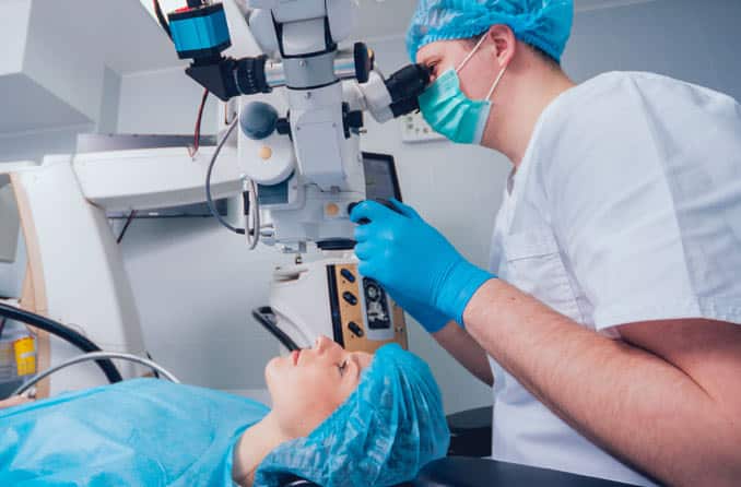
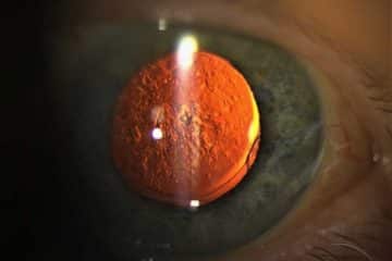
Laser cataract surgery, also known as refractive Laser-Assisted Cataract Surgery (ReLACS) involves the removal of the natural eye lens which has developed an opacification (cloudiness) known as cataract with the aid of a laser which provides a more reliable, repeatable, precise incision than a surgeon can do by hand, i.e traditional cataract surgery.
Laser cataract surgery is generally performed by an ophthalmologist in an ambulatory setting at a surgical center or hospital rather than an inpatient setting.
Laser cataract surgery helps to restore useful vision with a low complication rate.
More: Types of Cataract Surgery

Laser Cataract Surgery Procedure
The operation includes four major steps namely; the corneal incision, anterior capsulotomy, lens fragmentation and intraocular lens (IOL) implantation.
1. The corneal incision
The first step in cataract surgery is making an incision in the cornea.
In laser cataract surgery, the surgeon creates a precise surgical plan for the corneal incision with a sophisticated 3-D image of the eye called an OCT (optical coherence tomography).
The goal is to create an incision with a specific location, depth and length in all planes, and with the OCT image and a femtosecond laser, it can be performed exactly irrespective of the surgeons experience.
This is crucial in improving the chances of self-sealing after the procedure so as to reduce the risk of infection.
2. The capsulotomy
The eye’s natural lens is surrounded by a very thin, clear capsule. In cataract surgery, the front portion of the capsule is removed in a step called an anterior capsulotomy. This enables the surgeon to gain direct access to the cloudy lens (cataract).
It’s very important that the remainder of the lens capsule that remains intact in the eye is not damaged during cataract surgery, because it must hold the artificial lens implant in place for the rest of the patient’s life.
In laser cataract surgery, the anterior capsulotomy is performed with a femtosecond laser like the type used in LASIK vision correction surgery. Studies have shown that capsulotomies performed with a laser have greater accuracy and reproducibility compared to traditional cataract surgery.
3. Lens and cataract fragmentation
After the capsulotomy, the surgeon now has access to the cataract to remove it.
Laser cataract surgery oftens the cataract as it breaks it up. By breaking up the cataract into smaller, softer pieces, less energy should be needed to remove the cataract, so there should be less chance of burning and distorting the incision.
Laser cataract surgery may also reduce the risk of capsule breakage that can cause vision problems after surgery.
The lens capsule is as thin as cellophane wrap and it’s important that the the portion that is left inside the eye after cataract surgery is undamaged, so it can hold the IOL in the proper position for clear, undistorted vision.
4. Intraocular lens (IOL) implantation
After the removal of the cataract, an IOL is usually implanted into the eye, either through a small incision (1.8 mm to 2.8 mm) using a foldable IOL.
To reduce the need for prescription eyeglasses or reading glasses after cataract surgery, it is important that little or no astigmatism is present after implantation of presbyopia-correcting multifocal IOLs and accommodating IOLs. This can be achieved by making small incisions in the periphery of the more curved meridian; as the incisions heal, this meridian flattens slightly to give the cornea a rounder, more symmetrical shape.
Laser Cataract Surgery Risks and Complications
Complications after cataract surgery are relatively uncommon. However, as with every surgical procedure, there is risk of complications in laser cataract surgery. They include:
- Posterior vitreous detachment (PVD) – This does not directly threaten vision. PVD may be more problematic with younger patients, since many patients older than 60 have already gone through PVD. PVD may be accompanied by peripheral light flashes and increasing numbers of floaters.
- Retinal detachment – Retinal detachment normally occurs at a prevalence of 1 in 1,000 (0.1%), but patients who have had cataract surgery are at an increased risk (0.5–0.6%) of developing retinal detachment.
- Endophthalmitis – This refers to a a serious infection of the intraocular tissues, usually following intraocular surgery, or penetrating trauma.
- Edema – Swelling or edema of the central part of the retina, called macula, resulting in macular edema, can occur a few days or weeks after surgery. Most such cases can be successfully treated. Preventative use of nonsteroidal anti-inflammatory drugs has been reported to reduce the risk of macular edema to some extent
Other possible complications include: Swelling or edema of the cornea, sometimes associated with cloudy vision, which may be transient or permanent (pseudophakic bullous keratopathy). Displacement or dislocation of the intraocular lens implant may rarely occur. Unplanned high refractive error (either myopic or hypermetropic) may occur due to error in the ultrasonic biometry (measure of the length and the required intraocular lens power).
Laser Cataract Surgery Cost
Laser cataract surgery usually costs more than traditional cataract surgery, and the extra costs associated with laser cataract surgery typically are not covered by medical or health insurance.
The total cost for laser cataract surgery depends on a lot of factors such as the anesthetic fee, private hospital fee, private operating facility fee, extent of surgery required. The total cost of the procedure is around $3600 – $6000.
More: Oculoplastic Surgery


