Osteotomy is the surgical procedure whereby a bone is cut to shorten or lengthen it or to change its alignment. Le Fort Osteotomy refers to the group of osteotomies carried out to treat various degrees of maxillary fractures and deformities.
Maxillary fractures account for approximately 6-25% of all facial fractures.
Much of the understanding of patterns of fracture propagation in midface trauma originates from the work of René Le Fort. In 1901, he reported his work on cadaver skulls that were subjected to blunt forces of various magnitudes and directions. He concluded that predictable patterns of fractures follow certain types of injuries. Three predominant types were described.
Classification
- Le Fort I fractures (horizontal)
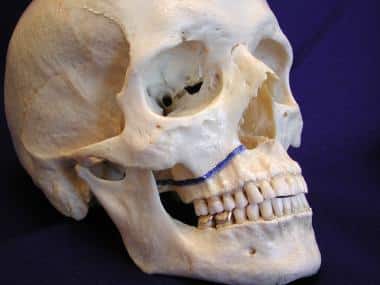
This may result from a force of injury directed low on the maxillary alveolar rim in a downward direction. The fracture extends from the nasal septum to the lateral pyriform rims, travels horizontally above the teeth apices, crosses below the zygomaticomaxillary junction, and traverses the pterygomaxillary junction to interrupt the pterygoid plates. See the image below.

Le Fort, I fracture
Signs and symptoms of Le Fort I fracture include slight swelling of the upper lip, abnormal alignment of the upper and lower teeth.
- Le Fort II fractures (pyramidal)
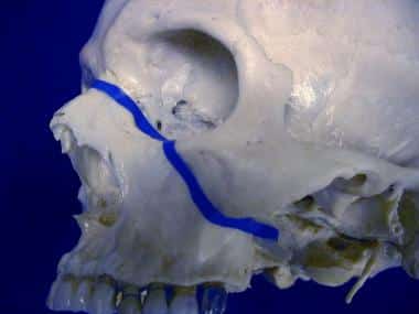
This may result from a blow to the lower or mid maxilla. Such a fracture has a pyramidal shape and extends from the nasal bridge at or below the nasofrontal suture through the frontal processes of the maxilla, inferolateral through the lacrimal bones and inferior orbital floor and rim through or near the inferior orbital foramen, and inferiorly through the anterior wall of the maxillary sinus; it then travels under the zygoma, across the pterygomaxillary fissure, and through the pterygoid plates. See the image below.

Le Fort II fracture
Signs and symptoms are step deformity at the infraorbital margin, mobile mid-face, anesthesia or paresthesia of the cheek.
- Le Fort III fractures (transverse)
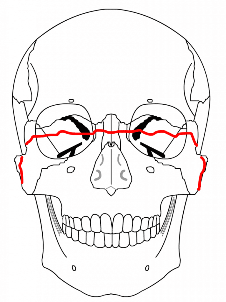
This is also termed craniofacial disjunctions, may follow impact to the nasal bridge or upper maxilla. These fractures start at the nasofrontal and front maxillary sutures and extend posteriorly along the medial wall of the orbit through the nasolacrimal groove and ethmoid bones. The thicker sphenoid bone posteriorly usually prevents the continuation of the fracture into the optic canal. Instead, the fracture continues along the floor of the orbit along the inferior orbital fissure and continues superolateral through the lateral orbital wall, through the zygomaticofrontal junction and the zygomatic arch. Intranasally, a branch of the fracture extends through the base of the perpendicular plate of the ethmoid, through the vomer, and the interface of the pterygoid plates to the base of the sphenoid. See the image below.

Le Fort III fracture
Signs and symptoms of Le Fort III fracture are tenderness and separation at the frontozygomatic suture, lengthening of face, and hooding of eyes.
Related: Types of Hip Fractures
Surgical Procedure for Le Fort Osteotomies
The Le Fort osteotomies are carried out under general anesthesia after the patient has been stabilized.
In Le Fort I osteotomy, the maxilla is osteotomised, down fractured and repositioned as required and then fixed using four adaptation plates; this is the classical approach described by Bell (1975).
Le Fort II osteotomy is performed through coronal and oral incisions because this gives the best access for the osteotomy, fixation and grafting and avoids further scars on the face. It is never possible to achieve the same degree of mobility as with the Le Fort I down fracture osteotomy.
When carrying out Le Fort II osteotomies, it is important to carefully consider the nasofrontal angle, which can become too obtuse, and the intercanthal distance, which can increase, with displacement anteriorly of the canthi.
The former can be avoided by judicious bone removal in order to reduce the nasofrontal angle at osteotomy. The latter often requires transnasal canthopexy through the osteotomy gap so that the ligaments can be approximated and moved into a more posterior position.
The Le Fort III Osteotomy is used to correct generalized growth failure of the midface involving the upper jaw nose and cheekbones (zygomas). The surgical approach and postoperative management are similar to the Le Fort II procedure.
Brow lift procedures may be carried out at the same time as Le Fort II and Le Fort III osteotomies.
Risks and Complications
As with any surgical procedure, Le Fort osteotomies have their risks of complications. They include:
Vascular complications
- Severe hemorrhage due to injury to the internal maxillary artery, the posterior superior alveolar artery, and the greater palatine artery
- Direct or indirect damage to major vessels in the neck or skull, including the internal carotid artery and internal jugular vein
- Thrombosis of the internal carotid artery due to the excessive head and neck extension
- Avascular necrosis of the maxilla
- Complications associated with prolonged hypotensive general anesthesia
Bony complication
- Nonunion or malunion of the maxilla due to poor skeletal fixation, insufficient bony contact, or instability of bone fragments
- Inadvertent fracture of the base of the skull
- Dysfunction of the temporomandibular joint
- Condylar head resorption
Dental complications
- Lingering malocclusion due to surgical relapse owing to strong soft tissue forces that antagonize surgical movements
- Anterior open bite in the early or late postoperative period
- Iatrogenic dental and periodontal injuries
- Soft tissue complications
- Oronasal communication during multi-piece maxillary orthognathic surgery
- Injury to the perioral soft tissues due to direct pressure, thermal burn, or sharp instrumentation
- Iatrogenic gingival/soft tissue pedicle injuries
Nerve complications
- Partial or total nerve injury to the infraorbital nerve with sensory loss to the upper lip, cheeks, and nose
- Paresthesia of sensory nerve supplies the maxillary teeth and mucosa
- Poor or unintended esthetic outcomes
- Alterations in the appearance of the midface soft tissues including widening of alar bases and changes in nasal tip position
- Shortening of upper lip vertical length
Recovery
All Le Fort osteotomies require an initial healing time of 2–6 weeks with secondary healing (complete bony union and bone remodeling) taking an additional 2–4 months.
The jaw is sometimes immobilized (movement restricted by wires or elastics) for approximately 1–4 weeks. However, the jaw will still require two to three months for proper healing.
Lastly, if screws were inserted in the jaw, the bone will typically grow over them during the two to three-month healing period.
Patients also may not drive or operate vehicles or large machinery during the consumption of painkillers, which are typically taken for six to eight days after the surgery, depending on the pain experienced.
Immediately after surgery, patients must adhere to certain infection prevention instructions such as daily cleaning, and the consumption of antibiotics.
Cleaning of the mouth should always be done regardless of surgery to ensure healthy, strong teeth. Patients can return to work 2–6 weeks after the surgery but must follow the specific rules for recovery for ~8 weeks.
Le Fort Osteotomies Cost
The cost of Le Fort osteotomies varies depending on the type of osteotomy to be performed with Level I being the cheapest and Level III the most expensive.
Also, the cost depends on whether other surgical procedures such as brow lift, are included with the osteotomy, anesthetic cost, private hospital fee, private operating facility fee.
The cost can run as low as $7,000 and as high as $20,000 or more.
More: Jaw Reduction Surgery


