Peroneal Tendon Subluxation Surgery
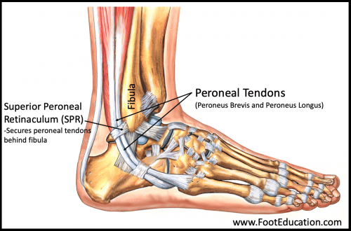
The peroneal tendons are anatomical structures that attach the peroneal muscles to the outer part of the foot.
One peroneal tendon attaches to the outer part of the midfoot, while the other tendon runs under the foot and attaches near the inside of the arch. The peroneal tendons function primarily to stabilize the foot and ankle and protect them from sprains.
The tendons are held in place by a retinaculum. Damage or injury to the structures that support the tendons may result in dislocation.
This condition is referred to as peroneal tendon subluxation. Treatment options for this condition may be surgical or non-surgical, depending on the severity and extent of the injury.
Anatomy

peroneal tendon subluxation
The peroneal muscles (Peroneus Brevis and Peroneus longus) are the primary muscles supporting the outer part of the ankle. The two muscles and their tendons are located along the outside of the fibula (lower leg bone) and cross behind the lateral malleolus (the outside ankle bone).
The tendons are stabilized by a superior peroneal retinaculum which is formed by the thickening of the superficial aponeurosis.
Causes
Peroneal tendon subluxation often results from a dorsiflexion force on the ankle with a rapid and strong contraction of the peroneal tendons. This leads to damage to the retraining reticulum causing the peroneal tendons to move out of place.
The injury to the retinaculum may be overlooked at first and there may be a differential diagnosis for ankle sprain. The result is that subluxation may begin much later. Hence a chronic problem results.
In the past, the sport most associated with this injury was alpine skiing, which correlates with the premise of forced dorsiflexion and acute muscle contraction against a fixed ankle in a ski boot.
However, recent literature does suggest that peroneal tendon subluxation can occur with those in several other sports, including football, soccer, basketball, baseball, softball and tennis.
More: Lipoma Removal Cost
Classification
Peroneal tendon subluxation can be classified into four (4) grades based on the Eckert and Davis classification.
Grade I. The retinaculum is stripped away from the fibula, resulting in the dislocation of the tendons.
Grade II. The fibrocartilaginous ridge and the retinaculum are avulsed from the posterior aspect of the fibula.
Grade III. Bony avulsion of the posterolateral aspect of the fibula containing the cartilaginous rim and a flake of bone permitting the tendon to slide beneath the periosteum.
Grade IV. The retinaculum is avulsed or ruptured from the posterior attachment, causing the retinaculum to be deep into the dislocating tendons.
Diagnoses
The diagnosis of peroneal subluxation begins with an examination of the ankle. The doctor will move your ankle in different positions to see when the tendons snap out of place and if they relocate.
One test involves holding pressure down on the ankle as you pull your foot up and out. During this test, the doctor feels behind the fibula to determine if the tendons are popping out of place.
If your doctor suspects a tear in the retinaculum, X-rays will probably be taken. X-rays can show if the torn retinaculum has pulled off a piece of the fibula bone.
This is called an avulsion fracture. X-rays are also used to look for other injuries to the ankle.
Treatment
Peroneal tendon subluxation can be treated through non-surgical and surgical means. The non-surgical means of treatment generally involve fewer risks, but it has high failure rates.
Surgery is often required when the conservative and non-surgical means are not successful.
Non-surgical Treatment
If the injury is acute, treatment without surgery may involve placing the ankle in a short-leg cast for four to six weeks. The goals are to allow the torn retinaculum to heal and to prevent chronic subluxation. Doctors may have their patients begin Physical Therapy once the cast is removed.
Your doctor may also prescribe medications. Anti-inflammatory medications can help ease pain and swelling and get you back to activity sooner.
These medications include common over-the-counter drugs such as ibuprofen.
Peroneal Tendon Subluxation Surgery
Surgery may be indicated to treat some grades of peroneal tendon subluxation. Various surgical techniques depend on the grade of the condition.
- Retinaculum Repair
Retinaculum repair is gaining popularity. This procedure restores the normal anatomy of the retinaculum that covers and reinforces the tendon sheath around the peroneal tendons.
In surgery to repair the retinaculum, the surgeon first makes an incision along the back and lower edge of the fibula bone. This lets the surgeon see the spot where the retinaculum is torn.
The surgeon uses a burr to create a trough along the fibula bone next to the original attachment of the retinaculum. The torn edge of the retinaculum is then pulled into the trough and sutured in place. The skin is closed with stitches.
- Groove Reconstruction
Groove reconstruction is done to deepen the groove so the personals stay in place behind the bottom tip of the fibula. In this procedure, the surgeon first makes an incision along the back and lower edge of the fibula bone.
The surgeon cuts a small flap in the bone near the bottom corner of the fibula. The surgeon then carefully folds the flap back like a hinge.
With the hinge held open, the doctor scoops out a small amount of bone under the flap to deepen the groove.
The surgeon closes the flap on its hinge and tamps it in place. A screw may be used to hold the flap down.
Next, the tendons are returned to their location behind the tip of the fibula. This procedure may also require repair of the retinaculum (see above). The skin is closed and sutured.
Risks and Complications
Like every surgical procedure, peroneal tendon surgery has risks and complications. They include:
- Bleeding
- Infection
iii. Recurrence of the peroneal tendon subluxation. The surgical repair can stretch out or be reinjured, leading to the recurrence of the peroneal tendon subluxation.
- Nerve injury. The sural nerve may be injured. This nerve supplies the sensation to the outside of the foot. An injury to this nerve can result in numbness over the outside of the foot or a burning sensation radiating toward the outside of the foot.
Peroneal Tendon Subluxation Surgery Cost
The total cost for peroneal tendon subluxation surgery depends on many factors, such as the anesthetic fee, private hospital fee, private operating facility fee, and the extent of surgery required. The total cost of the procedure is around $6500 – $7500.
Recovery
Recovery from surgery requires a moderately long period, usually between 2-6 weeks of immobilization, to allow the retinaculum and any bony procedures to heal.
This follows four to six weeks of fairly graduated and intensive rehabilitation. It is often six months or more before the patient has reached a lot of their improvement following the surgery.
More:


